



Polypoid Cystitis - Jessie’s Clinical Case
06 October 2026
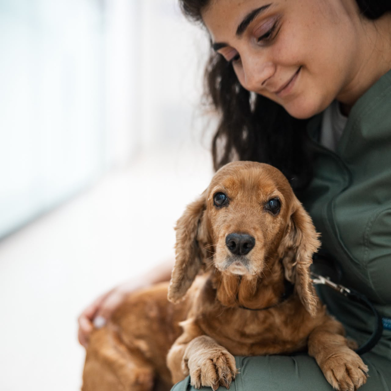
Jessie is an 8-year-old Cocker Spaniel who had experienced lower urinary tract signs for almost a year before coming to Veterinarium. These included haematuria (blood in the urine), pollakiuria (frequent urination) and dysuria (painful urination). Despite multiple courses of treatment during this period, her condition had not improved.
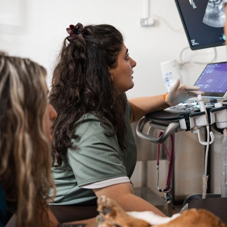
At our clinic, internal medicine veterinarian Dr. Nutsa Besashvili began the diagnostic work-up with an ultrasound examination of the urinary tract. This revealed marked, diffuse and asymmetric thickening of the bladder wall, with an irregular mucosal surface and polyp-like projections extending into the bladder lumen.
These ultrasound findings raised concerns about both chronic inflammation and possible neoplasia, prompting further investigation by cystoscopy. During the procedure, the bladder lining was examined and biopsy samples were collected for histopathological analysis. A urine sample was also obtained by cystocentesis for bacterial culture.
Histopathology revealed no evidence of neoplasia, while the urine culture yielded significant growth of Proteus spp.
Based on Jessie’s history, clinical signs and diagnostic findings, Dr. Nutsa Besashvili diagnosed polypoid cystitis.
Polypoid cystitis is a chronic inflammatory condition of the urinary bladder in which the lining thickens and develops polyp-like projections. In dogs, it is often associated with chronic bacterial urinary tract infections, with Proteus spp. among the commonly implicated bacteria.
Jessie’s treatment included a course of antibiotics selected according to antimicrobial susceptibility testing, anti-inflammatory and pain-relieving medication, and a urinary tract support supplement. She was also placed on a therapeutic urinary diet.
Soon after treatment began, Jessie’s urinary signs resolved completely, and she is now feeling very well.

Similar news

04 October 2026
Cocker Spaniel Lucy

03 October 2026
36th European Congress of Veterinary Internal Medicine for Companion Animals

26 September 2026
36th European Congress of Veterinary Internal Medicine for Companion Animals
23 September 2026
2-year-old mixed-breed dog Laima
13 September 2026